Man’s Brain Tumor Symptoms Were Actually Caused by Worms
▼ Summary
– A 60-year-old man in Spain had a worsening two-week headache, behavioral changes, and mild movement delay.
– CT scan showed multiple brain lesions with swelling; blood work revealed elevated IgE, suggesting allergies, autoimmune disease, or parasitic infection.
– Doctors initially suspected metastatic cancer, but whole-body scans and a colonoscopy found no malignancies.
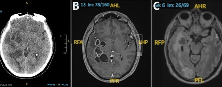
– MRI revealed the lesions were encapsulated tapeworm larvae (Taenia solium), not tumors.
– The man likely contracted the infection from coworkers who migrated from endemic regions, despite never traveling internationally.
A 60-year-old man in Spain sought medical care for a persistent headache that had lasted two weeks and was steadily worsening. He also reported subtle behavioral changes, prompting a thorough neurological examination.
During the exam, doctors observed a mild delay in his movements but found no other neurological deficits. His blood work was largely unremarkable, except for elevated IgE levels, a marker often associated with allergies, autoimmune conditions, or parasitic infections. A computed tomography (CT) scan of his head revealed more alarming findings: multiple lesions scattered across his brain, accompanied by swelling.
In a case report published in Emerging Infectious Diseases, the medical team described their diagnostic process. The patient was not immunocompromised and had never traveled internationally, leading them to initially suspect metastatic cancer as the most likely cause. To manage his headache, doctors prescribed an anti-inflammatory corticosteroid, which provided relief. They then launched an extensive search for a primary tumor, including a whole-body contrast-enhanced CT scan, a colonoscopy, and a hybrid positron emission tomography (PET)/CT scan commonly used to detect cancer. Yet none of these tests revealed any malignancies.
A follow-up brain scan using magnetic resonance imaging (MRI) offered clearer images of the lesions. This time, doctors could see that the spots were not tumors but encapsulated tapeworm larvae. On the MRI, the worms’ heads, known as scolexes, were visible, confirming the diagnosis.
This finding was unexpected, as tapeworms are not endemic to Spain, and the patient denied any travel history. However, the source of exposure may have been his former workplace. Until retiring a decade earlier, he had worked in construction, often alongside colleagues who had migrated from regions where pork tapeworms (Taenia solium) are common. These parasites spread through the fecal-oral route. Doctors speculate his infection was a rare case of cryptic transmission, likely from sharing meals and bathroom facilities with a coworker who unknowingly carried the tapeworm.
(Source: Ars Technica)