Brain Worm Infection Confirmed After Lab Eggs Grow Tails
▼ Summary
– A rice farmer in the Philippines experienced sudden uncontrollable shaking on one side of her body without prior warning symptoms.
– Doctors at the emergency department performed a physical exam that appeared normal but utilized MRI to reveal brain abnormalities.
– Imaging showed nodules surrounded by fluid in the left frontal lobe, which were identified as worm eggs causing neuroschistosomiasis.
– The infection is caused by Schistosoma parasitic worms, specifically S. japonicum, which is prevalent in parts of Asia including the Philippines and China.
– The parasite’s life cycle involves shedding eggs in waste, hatching into larvae, infecting aquatic snails, and eventually penetrating human skin to mature in the liver.
A rice farmer in the Philippines experienced a sudden, terrifying episode of involuntary movement that left half her body shaking for nearly a minute. The patient, who had previously appeared healthy with no signs of headache or weakness, rushed to the emergency department after the incident. Initial physical examinations yielded normal results, but advanced imaging told a different story. A magnetic resonance imaging (MRI) scan revealed a distinct cluster of bright nodules surrounded by fluid in her left frontal lobe. Doctors immediately suspected these were not tumors, but rather the eggs of a parasitic worm.
The diagnosis was confirmed as neuroschistosomiasis, a rare neurological complication caused by infection with Schistosoma parasites, commonly known as blood flukes. According to a case report published in the New England Journal of Medicine, the specific culprit was S. japonicum. This particular species is prevalent in the Philippines and China, as well as parts of Indonesia, despite its name suggesting a presence in Japan where it has actually been eliminated.
A Complex Parasitic Lifecycle
The infection stems from a biological cycle that is both intricate and relentless. The process begins when eggs are expelled from infected hosts, including humans and various animals, through urine or feces. Under favorable environmental conditions, these eggs hatch into free-swimming larvae. These microscopic travelers must locate specific species of aquatic snails, which they penetrate to undergo two developmental stages.
Once mature, the larvae emerge from the snail host with a distinctive forked tail. These tailed larvae, scientifically referred to as cercariae, possess the unique ability to burrow directly through intact human skin. Upon entry, they enter the vascular system and embark on a migratory journey through the lungs, heart, and finally the liver, where they mature into adult worms.
Adult Worms and Egg Production
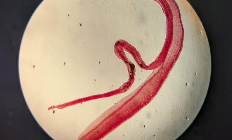
In their final stage, male and female worms pair up, with the slender female residing within a groove along the thicker male’s body. These paired adults typically migrate to the large blood vessels connecting the liver to the intestines. From this location, they continuously release eggs that eventually make their way into the host’s waste products, ready to be shed into the environment and perpetuate the cycle of infection.
(Source: Ars Technica)